I did some cool stuff last semester in my science classes that I’d like to show you guys.
The gist of it is… This picture:
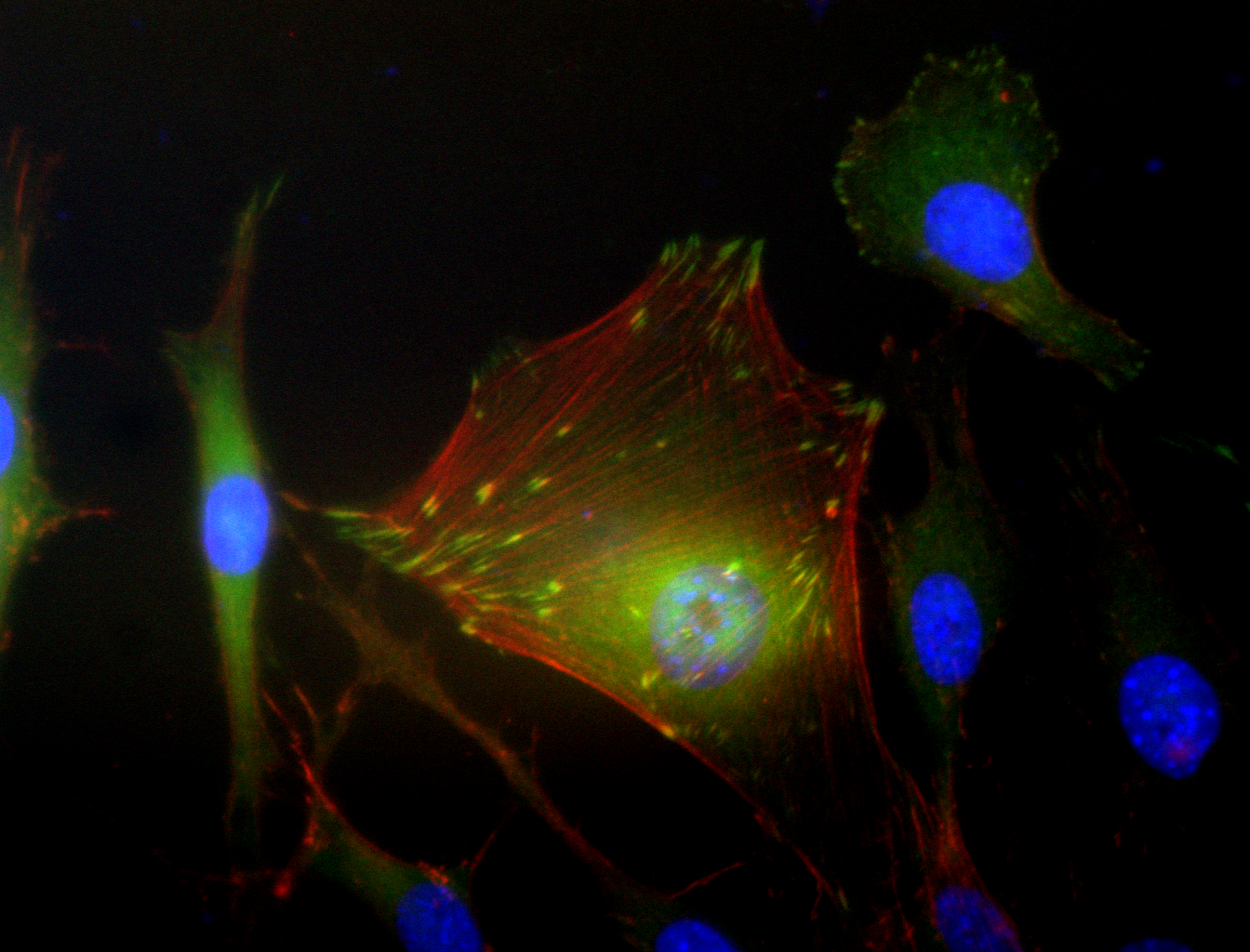
This is a picture taken by my lab group in my basic lab technique class last semester of a mouse fibroblast cell moving into a simulated wound on a glass slide.
Fibroblast cells are kind of like the contractors of your body when you get a scratch or wound. There are your first responders to the “disaster,” your immune system, and then fibroblasts go in to start the process of rebuilding your tissue by laying the foundation for other cells to move in.
A lot of scientists are interested in wound healing. How can we make it faster? How can we make it better so people don’t have lingering problems after the superficial injury has healed? How can we prevent infection? How can we prevent scarring?
Those questions are tested with a variety of experiments but one of the msot common is the scratch assay.
A bunch of fibroblasts are grown on a glass slide until they practically cover it. Then the slide is scratched.
The fibroblasts move into the scratch, thinking it is a wound. Their movement into the scratch is measured in a couple different ways and those measurements can tell us a little bit more about how wounds heal.
Which brings me back to the picture my lab group took. Obviously its got a lot of color and is very prety, but what are all those colors? What’s going on in that picture?
My lab group scratched the space above the big cell in teh picture. The cell is now moving into the scratch.
The red lines are called actin. Actin is the support structure of your cells. Cells move by extending actin filaments where they want to go and breaking them down behind them.
The green parts are called vinculin. Vinculin is spread throughout the cell and localizes into spots where the cell is attached to a surface to assist in adhereing to that surface. All those bright green spots are where the vinculin is helping the cell hold onto the glass slide.
The blue parts are cell nuclei. Each cell has one nucleus and I’ll bet you can pick out the one that belongs to all the actin and vinculin in the middle of this picture.
I did a lot more stuff on scratch assays in this class and leaarned a few new techniques, but the best part was definitely getting this picture.
Oh and apologies to any color blind people. I have no idea how to spearatae out the red and green things for you. Enjoy!
-GoCorral


Leave a comment